Ultrasound
Cooper Williams is one of a handful of veterinarians in the United States who is certified by the International Society of Equine Locomotor Pathology in advanced ultrasound imaging. Magda Stewart is working toward this certification as well and works closely with Dr. Williams advancing even further imaging expertise.
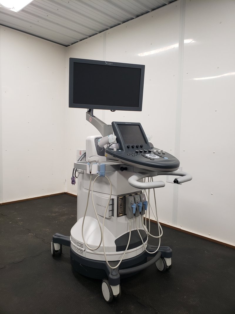
Ultrasound imaging is an invaluable tool for assessing soft tissues. Traditionally, equine veterinary ultrasound is used for diagnosing digital flexor tendon injuries and assessing reproductive status. With advanced imaging techniques an ultrasound exam allows us to diagnose and treat many musculoskeletal injuries that would only be found through MRI or scintigraphy. In addition we use ultrasound to guide needles directly into soft tissue lesions or for injecting joints and facets with complete accuracy such as sacroiliac or neck facet injections.
With advanced ultrasound techniques we can image the following areas:
- Distal front limb – foot, pastern, and fetlock joint
- Middle front limb – palmar fetlock, metacarpus and tendons, carpus, and carpal canal
- Proximal Front limb – forearm, elbow, and shoulder
- Distal hind limb – foot, pastern, fetlock, and metatarsus
- Middle hind limb – hock
- Proximal hind limb – stifle and thigh
- Neck and thoracolumbar area
- Lumbosacral area and pelvis